Home » Without Label » Chest Muscles Anatomy - 3d Chest Human Anatomy Or Anatomical And Muscle Set Or Collection Stock Illustration Illustration Of Medicine Abdominal 200786778 / The muscles of the chest and upper back occupy the thoracic region of the body inferior to the neck and superior to the abdominal region and include the muscles of the shoulders.
Chest Muscles Anatomy - 3d Chest Human Anatomy Or Anatomical And Muscle Set Or Collection Stock Illustration Illustration Of Medicine Abdominal 200786778 / The muscles of the chest and upper back occupy the thoracic region of the body inferior to the neck and superior to the abdominal region and include the muscles of the shoulders.
Chest Muscles Anatomy - 3d Chest Human Anatomy Or Anatomical And Muscle Set Or Collection Stock Illustration Illustration Of Medicine Abdominal 200786778 / The muscles of the chest and upper back occupy the thoracic region of the body inferior to the neck and superior to the abdominal region and include the muscles of the shoulders.. Chest muscles anatomy the chest is made up primarily of two muscles: Start with a pair of dumbbells extended above your chest. The chest is made up of two muscles: This muscle group is responsible for pushing movements and interacts synergistically with the anterior deltoid of the shoulder and. Moreover, the other muscular systems found inside the chest are digestive, circulatory and respiratory muscles which are not very noticeable, but they significantly have impact on complete human organism.
See chest anatomy stock video clips. All about the chest muscles the chest anatomy includes the pectoralis major, pectoralis minor and the serratus anterior. Anatomy chart courtesy of fcit the pecs attach to the humerus near the shoulder joint and originate on the breastbone in the center of the chest. It provides access to ct images in the axial plane, allowing the user to learn and review the lung anatomy interactively. This muscle group is responsible for pushing movements and interacts synergistically with the anterior deltoid of the shoulder and.
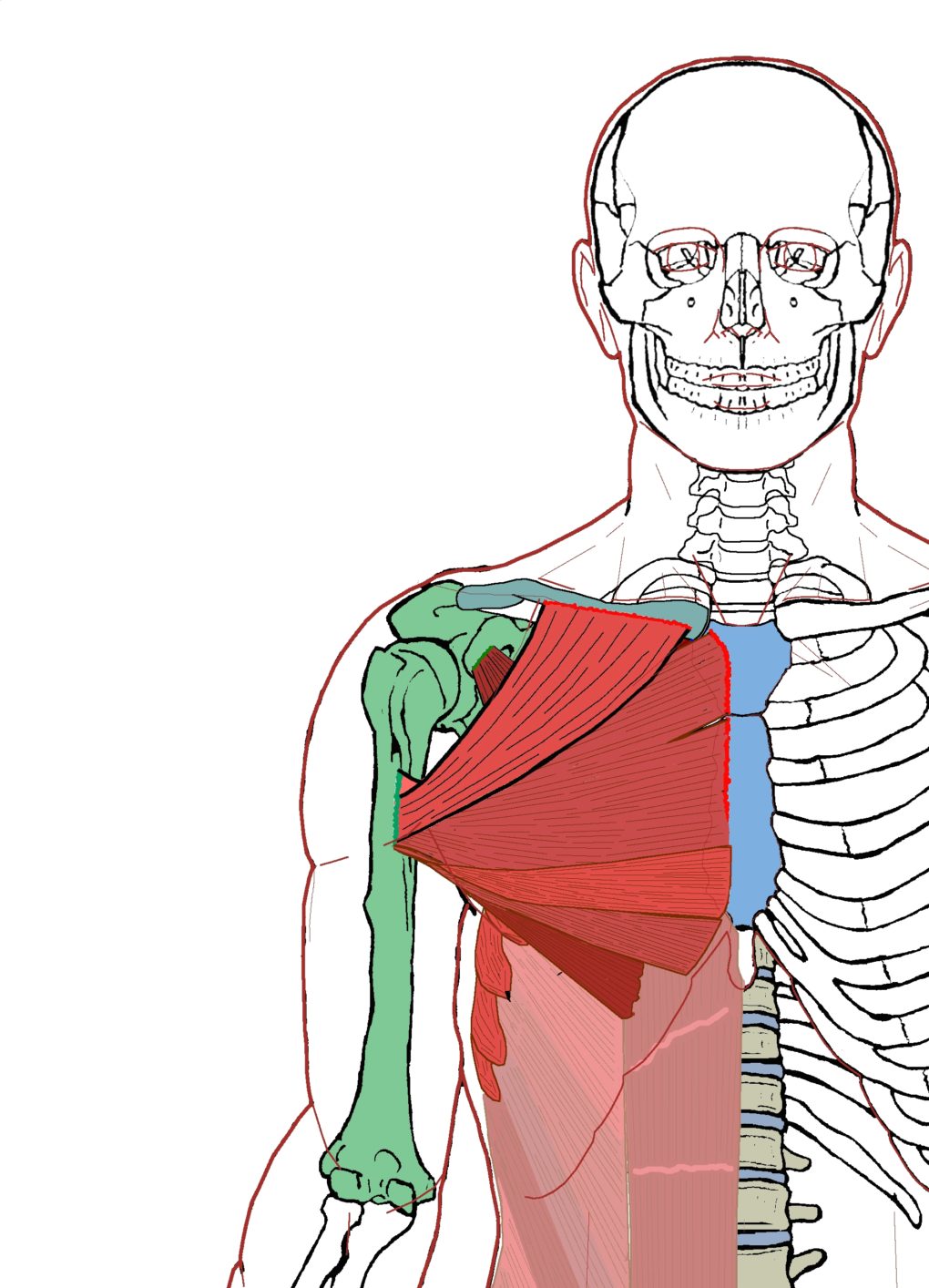
Extrinsic Chest Muscles Functional Anatomy Integrative Works from integrativeworks.com This muscle group is responsible for pushing movements and interacts synergistically with the anterior deltoid of the shoulder and. These heads are important to know because they can be specifically trained through particular movements. This mri chest (thorax) axial cross sectional anatomy tool is absolutely free to use. Anatomy chart courtesy of fcit the pecs attach to the humerus near the shoulder joint and originate on the breastbone in the center of the chest. Not everyone, however, has the chance to achieve stunning results when working on chest muscles. The thoracic wall is composed of the following muscles: The pectoralis major and the pectoralis minor, known collectively as your pecs. The pectoralis major, the larger and more superficial, originates at the clavicle (collarbone), the sternum, the ribs, and a tendinous extension of the external oblique abdominal muscle.
They then run across the front of the body and originate on the breastbone (picture a chicken breast).
The muscles of the chest and upper back occupy the thoracic region of the body inferior to the neck and superior to the abdominal region and include the muscles of the shoulders. See chest anatomy stock video clips. Chest muscles anatomy the chest is made up primarily of two muscles: Choose from 500 different sets of chest muscle anatomy flashcards on quizlet. The pecs are found attached to the humerus of the arm, right near where the shoulder joint is. This mri chest (thorax) axial cross sectional anatomy tool is absolutely free to use. To know whether or not an exercise targets the right muscles or not, scientists use a type of test called electromyography (emg). The chest or thorax is the region between the neck and diaphragm that encloses organs, such as the heart, lungs, esophagus, trachea, and thoracic diaphragm. Anatomy chart courtesy of fcit the pecs attach to the humerus near the shoulder joint and originate on the breastbone in the center of the chest. Applied anatomy of the chest wall and mediastinum petros mirilas michael e. However, our primary focus is on the chest's anatomy or the chest's main muscles in this section. There are two such muscles on each side of the sternum (breastbone) in the human body: System respiratory respiratory organs of human body digestive and respiratory system medical chest internal structure of human body medicine body lungs biology intestines stomach anatomy torso human internal.
Muscles the dominant muscle in the upper chest is the pectoralis major. It contains four muscles that exert a force on the upper limb: These heads are important to know because they can be specifically trained through particular movements. Pectoralis major, pectoralis minor, serratus anterior, and subclavius. Chest muscle anatomy the pectoralis major muscles (also known as the pecs) are located on the front of the rib cage, and form the major muscles of the chest.
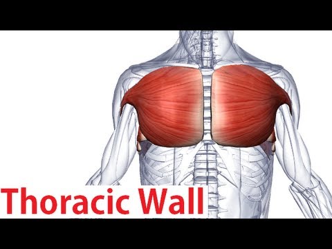
Muscles Of The Thoracic Wall Chest Muscles Anatomy Youtube from i.ytimg.com Beneath the pectoralis major is the pectoralis minor. The pectoralis major and the pectoralis minor, known collectively as your pecs. Here, we break down the anatomy of your chest muscles. Chest muscle anatomy the pectoralis major muscles also known as the pecs are located on the front of the rib cage and form the major muscles of the chest. While the minor muscle lay under the major muscle. Skandalakis chest wall embryogenesis the muscles of the chest develop from the somites found in the mesoderm. This video covers the definition, innervation and functions of the two pectoral muscles: Anatomy chart courtesy of fcit the pecs attach to the humerus near the shoulder joint and originate on the breastbone in the center of the chest.
Learn about each of these muscles, their locations, functional anatomy and exercises for them.
Pectoralis major and pectoralis minor. See chest muscles anatomy stock video clips. The chest is made up of two muscles: Applied anatomy of the chest wall and mediastinum petros mirilas michael e. This video covers the definition, innervation and functions of the two pectoral muscles: The chest and upper back muscles are known as deltoid, pectoral, teres (minor & major), latissimus dorsi and trapezius muscles. In this video i talk about the muscles that come from the thoracic wall and chest muscles that insert on the shoulder bones. This is one reason why there isn't a specific exercise just to build chest muscle. The thoracic wall is composed of the following muscles: This mri chest (thorax) axial cross sectional anatomy tool is absolutely free to use. Muscular anatomy of the chest pectoralis major. System respiratory respiratory organs of human body digestive and respiratory system medical chest internal structure of human body medicine body lungs biology intestines stomach anatomy torso human internal. Anatomy chart courtesy of fcit the pecs attach to the humerus near the shoulder joint and originate on the breastbone in the center of the chest.
The pectoralis major, the larger and more superficial, originates at the clavicle (collarbone), the sternum, the ribs, and a tendinous extension of the external oblique abdominal muscle. Use the mouse scroll wheel to move the images up and down alternatively use the tiny arrows on both side of the image to move the images. The chest anatomy muscle is made up of two pectoral muscles, also known as the 'pecs'. What you refer to as your chest is actually a group of called the pectorals. The chest is made up of two muscles:
Crosssection Anatomy Of Male Chest Abdomen And Groin Muscles Stock Illustration Getty Images from media.gettyimages.com What you refer to as your chest is actually a group of called the pectorals. The muscles of the chest and upper back occupy the thoracic region of the body inferior to the neck and superior to the abdominal region and include the muscles of the shoulders. These heads are important to know because they can be specifically trained through particular movements. The pectoralis major and the pectoralis minor, known collectively as your pecs. The chest or thorax is the region between the neck and diaphragm that encloses organs, such as the heart, lungs, esophagus, trachea, and thoracic diaphragm. Learn about each of these muscles, their locations, functional anatomy and exercises for them. Beneath the pectoralis major is the pectoralis minor. Applied anatomy of the chest wall and mediastinum petros mirilas michael e.
Pectoralis major, pectoralis minor, serratus anterior, and subclavius.
About the 6th week, the somites differentiate into the sclerotomes and the dermatomyotomes. All about the chest muscles the chest anatomy includes the pectoralis major, pectoralis minor and the serratus anterior. The pectoralis major, pectoralis minor, serratus anterior and subclavius. This is one reason why there isn't a specific exercise just to build chest muscle. The chest is made up of two muscles: The thoracic wall is composed of the following muscles: Beneath the pectoralis major is the pectoralis minor. The pectoral region is located on the anterior chest wall. Start with a pair of dumbbells extended above your chest. This page provides an overview of the chest muscle group. Moreover, the other muscular systems found inside the chest are digestive, circulatory and respiratory muscles which are not very noticeable, but they significantly have impact on complete human organism. See chest muscles anatomy stock video clips. The first muscle which is a major muscle has lower, middle and upper fibers.